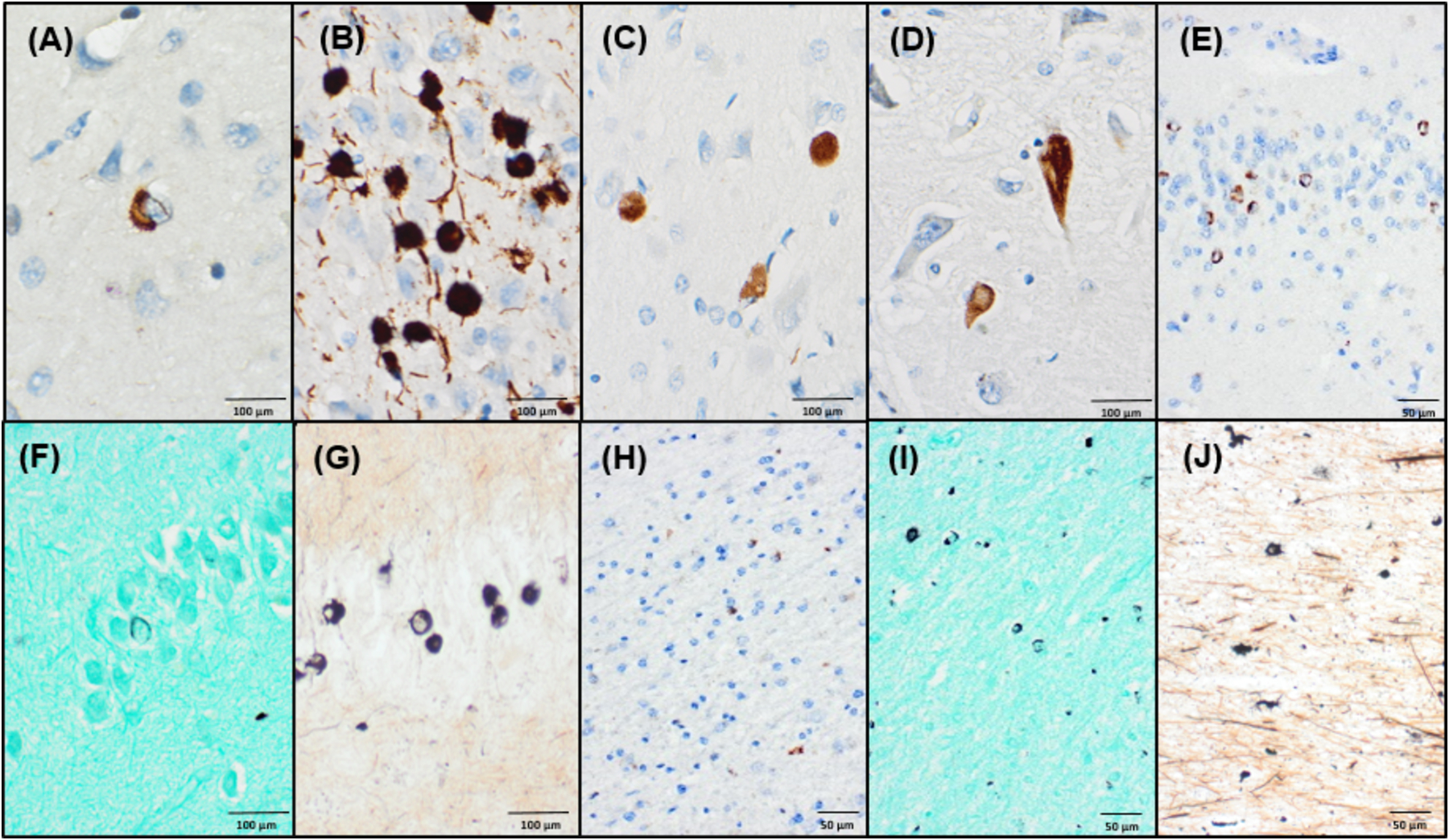
Figure 2. Immunohistochemical and histochemical characterization of limbic-predominant pathology in brain with longstanding MSA.

(A) α-synuclein (81A) positive neuronal cytoplasmic inclusion with a perinuclear skein in cingulate gyrus. (B) Tau (PHF-1) positive Pick-body like neuronal cytoplasmic inclusions seen in the posterior amygdala. (C) Many Pick-body like inclusions are also reactive for α-synuclein (81A). (D) α-synuclein (81A) positive neurofibrillary tangle in the CA1 region of the hippocampus. (E) Abundant α-synuclein (LB509) positive ring-like neuronal cytoplasmic inclusions are seen in the hippocampal dentate fascia. Only sparse ring-like inclusions are detected on Gallyas silver stain in the dentate fascia (F), compared to more abundant agyrophilic inclusions on Campbell-Switzer staining (G). (H-J) In frontal subcortical white matter, only sparse GCIs are detected by α-synuclein (LB509) IHC (H), but Gallyas staining (I) and Campbell-Switzer staining (J) more frequently highlight GCI pathology.
