
FIGURE 1.
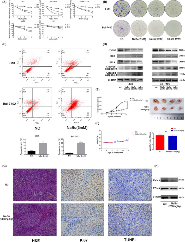
Effects of NaBu on proliferation and apoptosis of HCC in vitro and in vivo. (A) The CCK8 assay was performed to detect the viability of cells treated with NaBu (0–5 mM) for 24–72 h. (B) Colony formation by HCC‐LM3 and Bel‐7402 cells. (C) The apoptosis rate of HCC‐LM3 and Bel‐7402 cells after NaBu treatment for 48 h, determined by flow cytometry (n = 3, *p < 0.05 for NC vs. NaBu [3 mM]). (D) The protein expression of PCNA, Bax, Bcl‐2, cleaved caspase 3 and cleaved caspase 9, detected by Western blotting. (E) Tumour volume was recorded at the indicated time points (n = 4, *p < 0.05 for NC vs. NaBu). The gross manifestation of tumours. (F) Changes in mouse body weight were recorded at the indicated time points (n = 4, p > 0.05). (G) haematoxylin and eosin, TUNEL and Ki67 staining of tumour sections in the NC and NaBu groups (magnification 200×). (H) The protein expression of Bcl‐2 and PCNA in the mouse tumour tissues detected by Western blotting