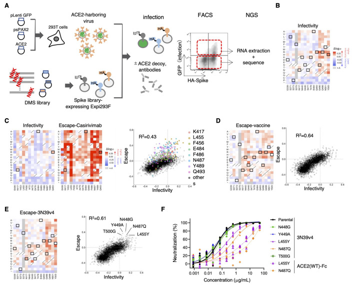
Fig. 6. Deep mutational scanning identified no single-residue mutation to induce complete escape from engineered ACE2.
(A) A schematic of the deep mutational scanning (DMS) approach to evaluate infectivity and escape from neutralizing agents is shown. FACS, fluorescence activated cell sorting. NGS, next generation sequencing. (B) Heatmaps illustrating how all single mutations that Omicron obtains affect its infectivity. Boxed squares indicate amino acid present in Omicron. Squares with a diagonal line through them indicate the original Wuhan strain amino acid. Heatmap squares are colored by mutational effect according to scale bars on the right. This and all subsequent coloring matches fig. S7 and S8. (C) The heatmaps show the alteration of infectivity and escape value from casirivimab in casirivimab antigen-binding sites of the spike protein. Boxed squares are mutations found in Omicron (left). Correlation in mutation effects on infectivity and escape from casirivimab are shown on the right. Colored dots are all amino acid substitution at the indicated major casirivimab antigen-binding sites. (D) The heatmap illustrates how all single mutations that Omicron obtained affect its escape from vaccinated serum samples. Boxed squares indicate amino acid present in Omicron (left). Correlations in mutation effects on infectivity and escape from vaccinated serum are shown on the right. (E) The heatmap illustrates how all single mutations that Omicron obtained affect its escape from 3N39v4. Boxed squares indicate amino acid present in Omicron (left). The dot plot shows the correlation between Omicron infectivity and escape from 3N39v4. Indicated mutants were individually analyzed in (F). (F) Neutralization efficacy was measured in 293T/ACE2 cells for 3N39v4 or wild type ACE2 decoys against the five indicated escape candidates. n = 4 technical replicates.

