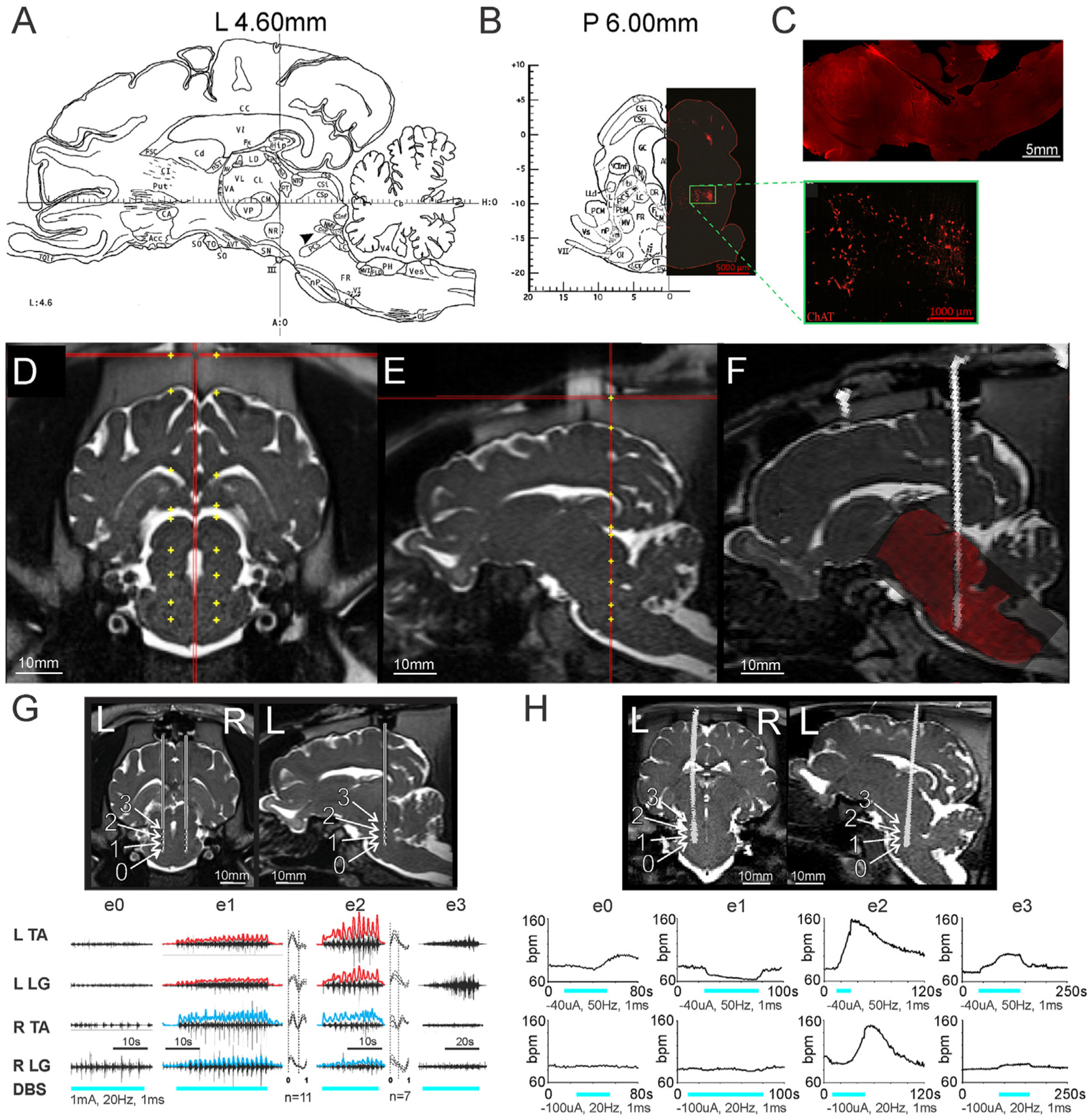
Fig. 1. Intraoperative stimulation of the MLR elicits locomotor-like EMG activity in the anesthetized pig.

(A) Sagittal slice from Félix et al. [22], at 4.6 mm lateral to midline depicts the location of the cuneiform nucleus (arrowhead, CnF), slightly inferior and anterior to the inferior colliculus. (B) Left: coronal slice from Félix et al. [22], at 6 mm posterior to the anterior limit of the posterior commissure at midline. Right: ChAT immunohistochemistry of a matched coronal slice, depicting a cluster of cholinergic neurons (green box, inset), traditionally defining the pedunculopontine nucleus, which is located ventral to the CnF. (C) ChAT immunohistochemistry of a sagittal brainstem slice similar to the atlas view in (A), showing the cholinergic cluster ventral to electrode track. (D) Coronal and (E) sagittal views of example planned electrode trajectories targeting the MLR on pre-operative MRI. (F) Example of pre-op MRI/post-op CT fusion, with superimposition of post-mortem histology confirming electrode position via track. (G) Top: coronal view, bilateral electrode placement and sagittal section (left side) showing calculated positions of electrodes 0–3 (e0, e1, e2, e3). Positions are approximations of the electrode size overlaid on the MRI cannula trajectory based on depth advanced. Bottom: EMG responses to stimulation of electrodes 0–3 on left side. Rectified and high pass filtered (>2Hz) traces of individual EMG traces from e1 and e2 are overlaid in red (left) and blue (right). Step cycle averages for e1 and e2 are shown on right of each muscle, with the number of step cycles averaged indicated. Repetitive EMG activity is observed with e1 and e2 stimulation, located within the cuneiform and adjacent subcuneiform region. EMG gain for each muscle is constant between trials. (H) Top: Coronal and sagittal views showing electrode position on CT scan superimposed onto brain MRI of another animal. Bottom: Heart rate responses to stimulation at each electrode position, using 20Hz or 50Hz stimulation. R: right, L: left, ECR: extensor carpi radialis; FCU: flexor carpi ulnaris; LG: lateral gastrocnemius, TA: tibialis anterior, SCL: step cycle length. For other atlas abbreviations please see Ref. [22]. G: From Noga et al. [18], with permission.
