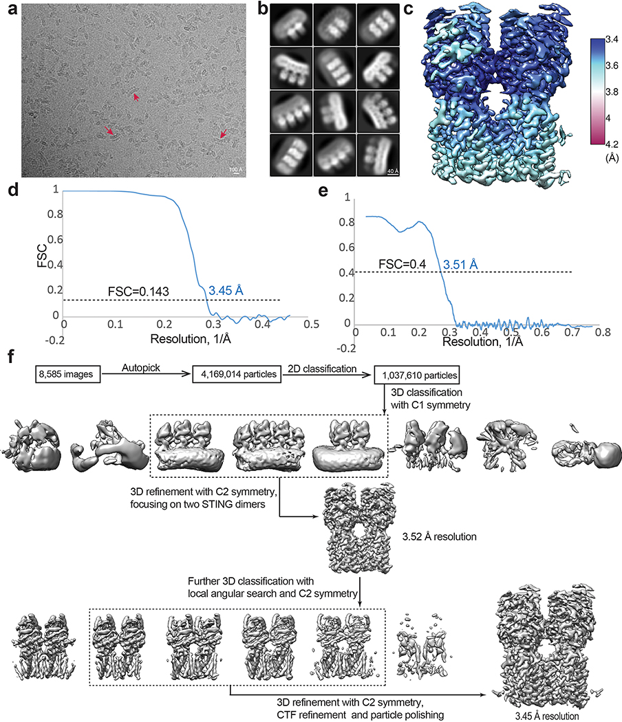
Extended Data Fig. 2. Image processing procedure of human STING tetramer bound to both cGAMP and C53.
a, Motion corrected micrograph. Red arrows highlight high-order oligomers of STING. The curved overall shape of the oligomers is evident from these examples. b, 2D class averages of high-order oligomers of human STING. Large oligomers were segmented into particles containing four dimers at the maximum. c, Final 3D reconstruction of the tetramer colored based on local resolution. d, Gold-standard FSC curve of the final 3D reconstruction. e, FSC between the final map and the atomic model. f, Image processing procedure.

