
Figure 3: Visualizing the misfolded oligomers using the AggTag method.
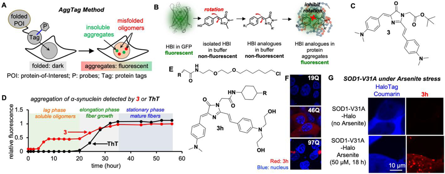
(A) Overview of the AggTag method. (B) Chemical modulation of the FP chromophore (HBI) to detect protein aggregation. (C) Chemical structure of 3. (D) 3 detects misfolded oligomers during α-synuclein aggregation. (E) Chemical structure of 3h. (F–G) Detecting misfolded oligomers of Htt (F) and SOD1-V31A (G). Reproduced with permission from Ref. 2. Copyright 2018 American Chemical Society.