Figure 3.

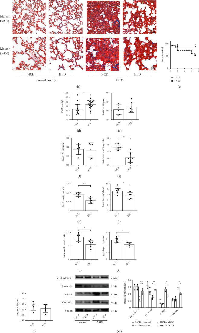
High-fat diet-induced obesity protects against ARDS by promoting pulmonary endothelial barrier and attenuating pathological fibroproliferation in mice. Histopathologic alterations of lung tissue by hematoxylin and eosin (H&E) staining (a) and Masson's trichrome staining (b) in the normal chow diet (NCD) induced lean control mice and high-fat diet (HFD) induced obese mice under normal and ARDS conditions (Scale bar = 50 μm for magnification ×200 and scale bar = 20 μm for magnification ×400). Representative images are shown from three replicated independent experiments. Comparison of mortality (c), PaO2 (d), the level of IL-6 (e), TNF-α (f), MMP9 (g), and protein (h) in bronchoalveolar lavage fluid (BALF), Evans blue-dyed albumin (EBDA) extravasation (i), wet/dry ratio (j), and collagen content (k) and TGF-β concentration in lung homogenate (l) between the NCD and HFD fed mice with ARDS. n = 16 mice per group (c). n = 8 mice for NCD group, and n = 13 mice for HFD group (d). n = 6 mice per group (e)–(g). n = 5 mice for the other group. (m) Western blot analysis of the protein expression of VE-cadherin, β-catenin, α-SMA, and Vimentin in the lung tissue. (n) Western blot analysis of the protein expression of TGF-β and TGFβR1 in the lung tissue. Relative abundances of protein bands were normalized to β-actin as shown in the bar graphs. Relative phosphorylation levels of protein are expressed normalized to the corresponding total protein. n = 3 mice per group analyzed in three replicated independent experiments. Data are presented as mean ± S.D. Significant differences are shown by ∗P < 0.05, ∗∗P < 0.01, and ∗∗∗P < 0.001.
