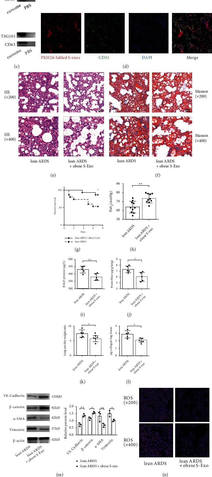
Figure 4.

Obesity promotes pulmonary endothelial barrier and attenuates pathological fibroproliferation and oxidative stress via circulating exosomes. (a) Transmission electron microscope (TEM) analysis of serum exosomes from obese or lean control mice. Scale bar = 200 nm (left panel) and 100 nm (right panel). (b) Particle size and concentration of exosomes were detected by nanoparticle tracking analysis (NTA). (c) Exosome-specific marker TSG101 and extracellular vesicle-related protein marker CD63 were verified by western blot analysis. (d) PKH-26 dye labeled exosomes were intravenously administered to recipient mice. The efficient uptake of exosomes was confirmed by the appearance of red fluorescent PKH-26 dye in pulmonary capillaries, which were labeled with the endothelial marker CD31 (green fluorescence). Scale bar = 50 μm. Histopathologic alterations of lung tissue by H&E (e) and Masson's trichrome staining (f) in lean mice with ARDS, which were pretreated with exosomes from obese mice serum (100 μg/mL in a total volume of 300 μL of PBS every week for three weeks). Representative images are shown from three replicated independent experiments. The mortality (g), PaO2 (h), BALF protein (i), EBDA extravasation (j), wet/dry ratio (k), and collagen content (l) in lean mice subjected to ARDS and those pretreated with exosomes from obese mice serum. n = 15 mice per group (g). n = 10 mice per group (h). n = 5 mice for the other group. (m) Western blot analysis of the protein expression of VE-cadherin, β-catenin, α-SMA, and Vimentin in the lung tissue. Relative abundances of protein bands were normalized to β-actin as shown in the bar graphs. n = 3 mice per group analyzed in three replicated independent experiments. (n) ROS production and (o) free GSH concentration in lung tissue.
