
Figure 8.
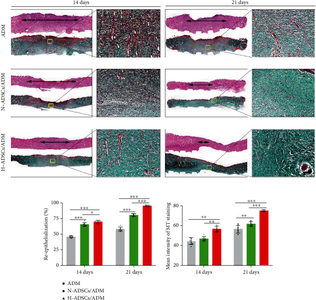
H-ADSCs/ADM facilitating reepithelialization and collagen deposition in diabetic wounds. (a) The images of H&E- and MT-stained sections in the ADM, N-ADSCs/ADM, and H-ADSCs/ADM groups at 14 or 21 days after the operation. “Double-headed black arrows” mean the edges of the scars. Bar = 100 μm. (b) Quantitative analysis of the reepithelialization extent and the mean intensity of MT staining in the histological sections of the ADM, N-ADSCs/ADM, and H-ADSCs/ADM groups, n = 8 per group. All data are shown as means ± standard deviation (∗P < 0.05, ∗∗P < 0.01, and ∗∗∗P < 0.001).