
Figure 9.
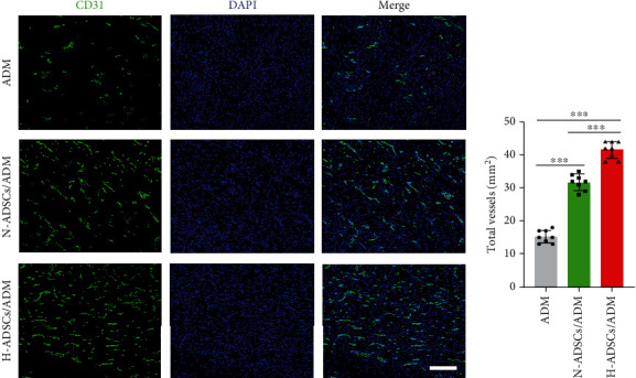
H-ADSCs/ADM membrane stimulating angiogenesis in diabetic wounds. (a) Immunofluorescence analysis of sections for CD31 showed the formation of microvessels in the ADM, N-ADSCs/ADM, and H-ADSCs/ADM groups. Bar = 100 μm. (b) CD31 area quantification showed the quantitative analysis of the number of total blood vessels in wounds at day 7 postoperation, n = 8 per group. All data are shown as means ± standard deviation (∗P < 0.05, ∗∗P < 0.01, and ∗∗∗P < 0.001).