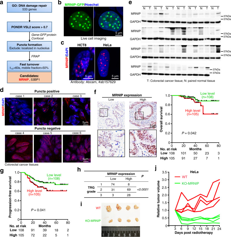
Fig. 1. MRNIP formed puncta in tumor cells and its high expression was associated with the radioresistance and poor prognosis of CRC patients.
a Screening of DNA damage repair-related proteins that undergo LLPS. Details could be found in Supplementary Figure 1a. b MRNIP-GFP showed puncta in the nucleus of HEK 293 T cells. HEK 293 T cells were transfected with plasmid for 24 h before observation. c Immunofluorescence of endogenous MRNIP in HCT8 and HeLa cells. d Immunofluorescence assay showing that MRNIP formed puncta in 3 out of 6 CRC tissues. Frozen sections of CRC tissues were analyzed using Immunofluorescence assay. e MRNIP protein was upregulated in CRC tissues. Twenty-seven CRC tissues and adjacent normal tissues were analyzed. f, g Higher MRNIP expression was correlated with shorter survival time of CRC patients. The correlations were analyzed with Kaplan-Meier curve and Log-rank test. h CRC patients with higher MRNIP level were more resistant to radiotherapy. TRG, Tumor regression grade. For (f–h), CRC tissues from 213 patients were analyzed. i, j Xenograft model showed that MRNIP depletion sensitized tumor cells to radiation. For (j), each line represents one xenograft.

