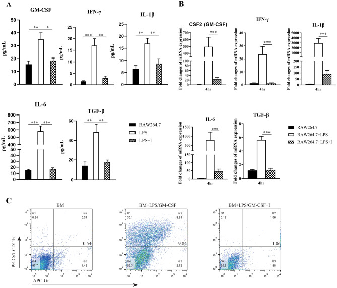
Fig. 4.
MyD88 inhibitor administration suppressed the differentiation of myeloid cells into MDSCs in vitro. Supernatant concentrations detected by ELISA (A) and relative levels of mRNA transcripts detected by RT-qPCR (B) of GM-CSF, IFN-γ, IL-1β, IL-6 and TGF-β1 in RAW 264.7 cells are shown. Data are expressed as the mean ± SD of each group from three independent experiments. Data from the RT-qPCR were normalized to unstimulated cells. *P < 0.05; **P < 0.01; ***P < 0.001. C) BM cells were cultured in the present of GM-CSF and LPS for eight days and a phenotypic analysis on induced CD11b+Gr-1+ MDSCs was performed by flow cytometry. Groups: the unstimulated, LPS or LPS/GM-CSF-stimulated and MyD88 inhibitor-treated (I) cells groups

