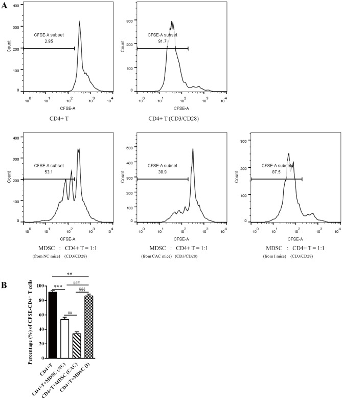
Fig. 5.
The suppressive capacity of CD11b+Gr-1+ MDSCs on the proliferation of activated CD4+ T cells was reserved by MyD88 inhibitor administration. MDSCs derived from the normal control, CAC and MyD88 inhibitor-treated (I) mice were co-cultured with CFSE-labeled autologous CD4+ T cells in proportion (1:1) in the present of anti-CD3/anti-CD28-antibody-coated microbeads. A) CFSE-labeled T cells were analyzed by flow cytometry for proliferation. B) Percentage of CFSE-labeled CD4+ T cells were stimulated by anti-CD3/anti-CD28-antibodies. Data were from three independent experiments. Values are means ± SD. ***P < 0.001, **P < 0.01 vs. stimulated CD4+ T cells; ###P <0.001, ##P < 0.01 vs. stimulated CD4+ T cells co-cultured with MDSCs from NC mice; §§§P <0.001 vs. stimulated CD4+ T cells co-cultured with MDSCs from CAC mice

