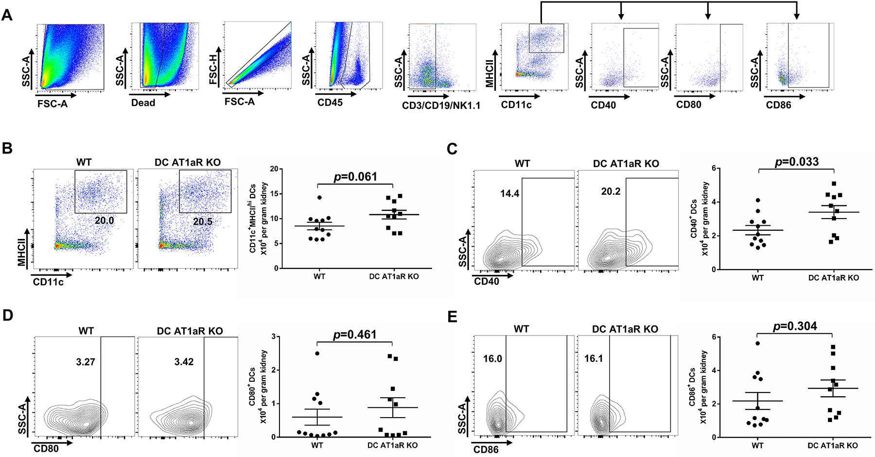
Figure 4. AT1aR suppresses renal dendritic cell (DC) activation during hypertension.

Flow cytometric assessment of co-stimulatory molecule expression on CD11c+MHCIIhi DCs isolated from the kidney after 4 weeks of Ang II infusion. (A) Gating strategy for parsing for CD40, CD80, or CD86 positivity on DCs in the kidney, based on “fluorescence minus one” strategy. (B) Representative flow plots and the absolute number of CD11c+MHCIIhi Cells from WT and DC AT1aR KO groups. (C-E) Representative flow plots and the absolute number of (C) CD40+, (D) CD80+, and (E) CD86+ DCs from WT and DC AT1aR KO mice. N≥10 mice/group. Data are mean ± SE.
