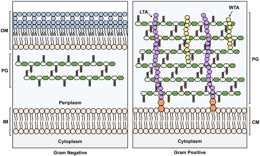
Figure 1. Architecture of the Gram-negative and Gram-positive cell envelopes.

The Gram-negative inner membrane (IM) and the Gram-positive cytoplasmic membrane (CM) are phospholipid bilayers. In contrast, the outer membrane (OM) of Gram-negative bacteria is asymmetrical with phospholipids in the inner leaflet and lipopolysaccharide (LPS, with sugars in blue) in the outer leaflet. IM: inner membrane, PG: peptidoglycan, OM: outer membrane, LTA: lipoteichoic acid, WTA: wall teichoic acid. In the PG cartoon structure, the glycan strands are represented as polymers of hexagons, with N-acetylglucosamine (GlcNAc) in dark green and N-acetylmuramic acid (MurNAc) in light green, while the stem pentapeptides are shown as light orange, blue, purple and dark orange spheres. For more details on the structure of the PG building block, refer to Figure 2 and Section 2.1.
