Figure 7.
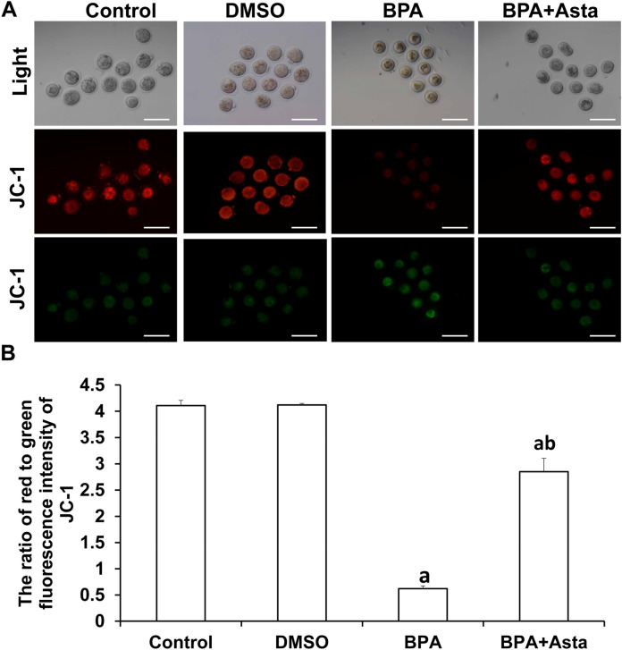
Analysis of mitochondrial membrane potential in oocytes. (A) JC-1 probe was used to detect the mitochondrial membrane potential in oocytes. JC-1 forms J-aggregates and produces red fluorescence under normal mitochondrial membrane potential (Scale bar, 200 μm). Under reduced or lost mitochondrial membrane potential, JC-1 exists in J-monomers and produces green fluorescence. (B) The average fluorescence intensity was analyzed in oocytes (N = 10). Compared with control, aP < 0.05; and compared with BPA group, bP < 0.05.

