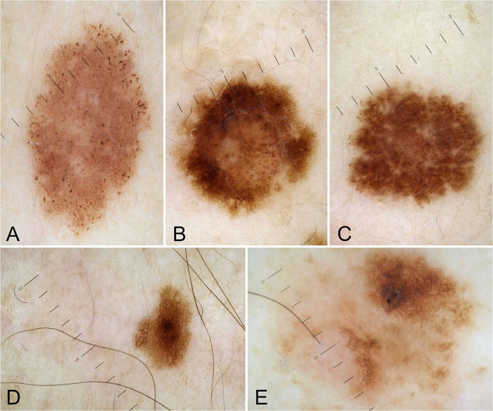
Figure 1.
Dermoscopic patterns of nevi: uniform globular pattern (A); mixed pattern composed of a central globular or structureless area surrounded by a network (B); mixed pattern composed of a central network or structureless brown-gray area surrounded by a peripheral rim of small brown globules (C); reticular pattern (D); unspecified pattern (E).

