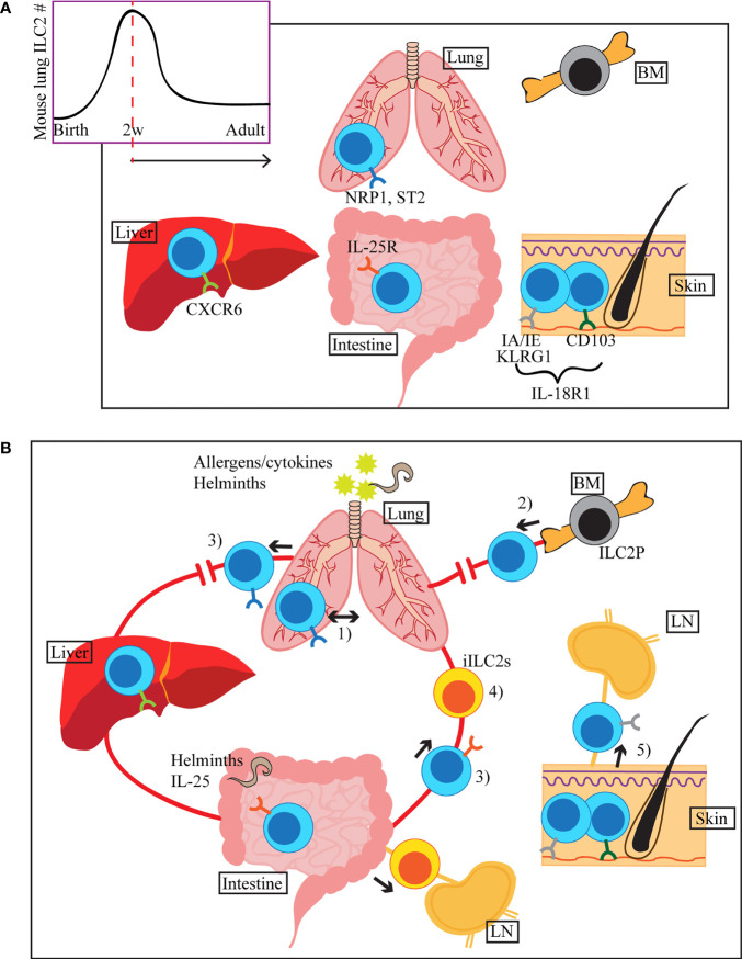
Figure 1.
(A) ILC2s seed tissues early during the neonatal period, with a peak of ILC2 accumulation around 2 weeks after birth (mouse lung). Once in tissues, they receive environmental cues and adapt tissue specific phenotypes, indicated by different colours of the surface receptors in the figure. Common ILC2 markers in each tissue are shown, but they can also be expressed in other organs. (B) ILC2s migrate within the lung upon i.n. IL-33 or Aa treatment (1). During allergen-induced respiratory inflammation or tissue disruption, ILC2s are recruited from hematogenous sources (2). When activated by allergens/cytokines or helminth infection, some of the lung and small intestinal ILC2s appear in the peripheral blood (3). Stimulation by i.n. IL-33 or papain induces migration of a subset of lung ILC2s to the liver, where they contribute to the regulation of local immunity (3). Upon helminth infection or i.p. IL-25 stimulation, small intestine-derived inflammatory ILC2s (iILC2s) appear in circulation and accumulate in the lung, liver, mesenteric LN and spleen (4). iILC2s ultimately become conventional ILC2s in the lung. Tissue resident and circulating ILC2s are present in the skin. During atopic dermatitis-like inflammation, circulatory ILC2s migrate to the draining LN (5).

