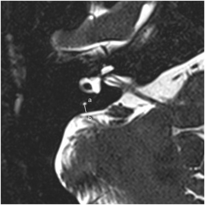
Figure 1.

A 0.5-mm axial 3D-SPACE MRI scan showing detailed image of the right ear at the level of the measured distance between the vertical part of the posterior semicircular canal (a) and the posterior fossa (b). 3D-SPACE, three-dimensional sampling perfection with application optimized contrasts using different flip angle evolutions.
