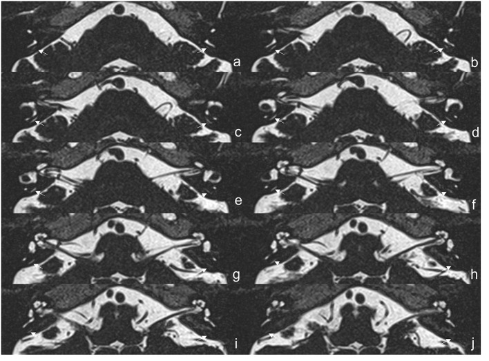
Figure 2.
The 3D-SPACE MRI images of a 57-year-old female with vestibular migraine (VM). (a–j) Axial, high-resolution, and T2-weighted MRI scan showing visualization of the vestibular aqueduct on both sides. 3D-SPACE, three-dimensional sampling perfection with application optimized contrasts using different flip angle evolutions, VM, vestibular migraine.

