FIGURE 1.
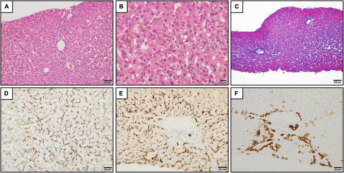
Histopathologic findings in the liver. (A) Liver tissue showed no significant lobular architecture (hematoxylin and eosin staining). Scale bar represents 50 μm. (B) Cholestasis was observed in the hepatocytes, Kupffer cells, and bile canaliculi at zones 2 and 3 (hematoxylin and eosin staining). Scale bar represents 20 μm. (C) There was no significant fibrosis (Azan staining). Scale bar represents 100 μm. (D–F) Immunohistochemistry for bile salt export pump (D), CD10 (E), and keratin 7 (F). The strong expression of bile salt export pump and CD10 were observed at the canalicular membrane (D,E). The keratin 7 expression was found in the hepatocytes in the periportal areas. The intralobular bile duct arrangement was preserved (F). Scale bars represent 50 μm.

