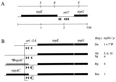
FIG. 1.
Linearized physical maps of B. aphidicola trpEG plasmids. All genes are transcribed in the rightward direction. Open arrowheads indicate the approximate position and orientation of DnaA boxes. (A) Restriction site and genetic map of pBTc2 from B. aphidicola (T. caerulescens). The region denoted ori? contains the putative origin of replication. Restriction enzyme sites: X, XbaI; B, BglII; E, EcoRI. (B) Genetic maps of the repeated units of B. aphidicola (Aphididae) trpEG plasmids (adapted from reference 42 with permission from the publisher). B(sp.), species names (see below); trpEG/p, number of repeat units per plasmid (42); ori-3.6, putative origin of replication; Ψ, repeat units containing trpEG pseudogenes (32); striped box and ΨrepAC, location of a putative repAC pseudogene; open rectangle, 19-bp element similar to consensus sequence of RepA/C iterons. Species abbreviations: Dn, Diuraphis noxia; Ap, Acyrthosiphon piseum; Sg, Schizaphis graminum; Rp, Rhopalosiphum padi; Rm, Rhopalosiphum maidis.

