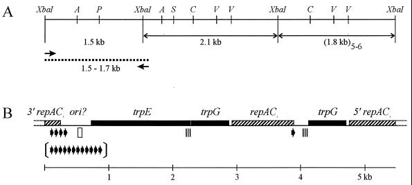
FIG. 4.
Physical map of cloned and sequenced portion of pBPs2 from B. aphidicola (P. spyrothecae). (A) Restriction site and clone map. Double-headed arrows, cloned XbaI restriction fragments; arrows, oligonucleotide primers used for PCR; dotted line, cloned PCR product. Restriction enzyme sites: A, AccI; P, PvuII; S, SacI; C, ClaI; V, EcoRV. (B) Genetic map. All genes are transcribed in the rightward direction. The region denoted ori? contains the putative origin of replication. Arrows, 19-bp iteron unique to pBPs2; arrows between brackets illustrate the copy number of the same iteron in the 1.70-kb PCR fragment; open rectangle, 19-bp element differing in one nucleotide from consensus sequence of RepA/C iterons; triple strip, 129-bp sequence identical to the 3′ end of trpE.

