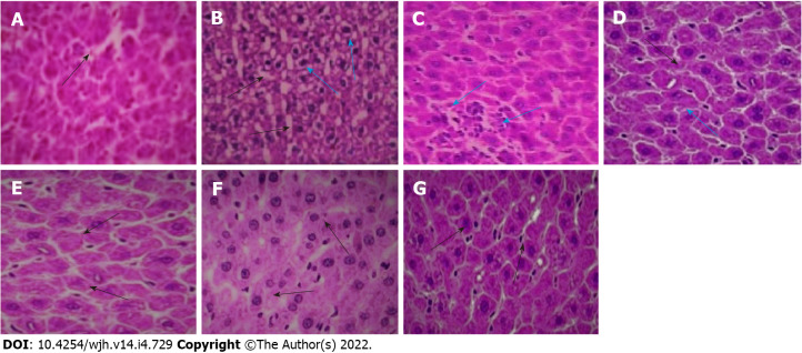
Figure 6.
Histological sections of rat liver. A: Normal control (the black arrow shows normal intercellular spaces and negligible vacuolation); B: Fluoxetine (10 mg/kg) treated (the black arrows show increased vacuolation and cellular degeneration, blue arrows indicate presence of inflammatory cells); C: Fluoxetine + baicalin (50 mg/kg) treated (blue arrows indicate a patch of inflammatory cell infiltration); D: Fluoxetine + baicalin (100 mg/kg) treated (the black arrow depicts regular cellular pattern and normal sinusoidal space, blue arrow indicates fewer inflammatory cells); E: Fluoxetine + silymarin (100 mg/kg) treated (the black arrows show normal cells and intercellular spaces similar to normal control); F: Baicalin (100 mg/kg) treated (the black arrows show normal cellular structure); G: Silymarin (100 mg/kg) treated (the black arrows depict normal cells and intercellular spaces).

