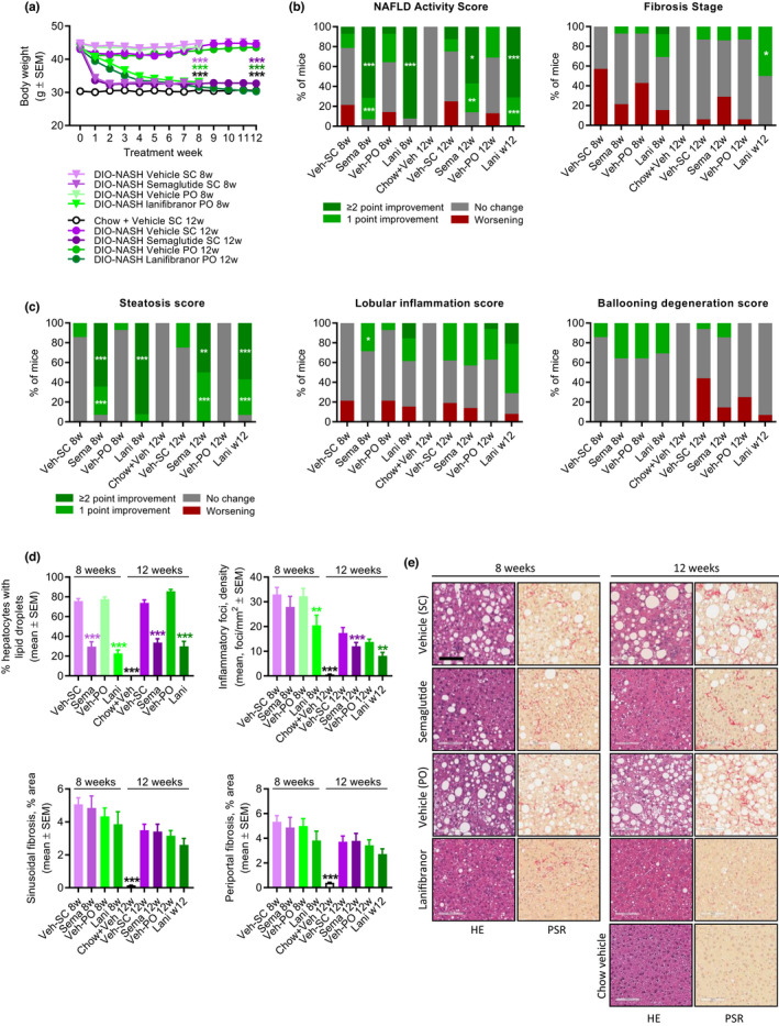
FIGURE 5.

Semaglutide and lanifibranor differentially improves liver histopathological hallmarks in GAN DIO‐NASH mice. GAN DIO‐NASH mice were administered (q.d.) semaglutide (Sema, 30 nmol/kg, s.c.), lanifibranor (Lani, 30 mg/kg, p.o.), or corresponding vehicle (Veh, s.c., or p.o.) for 8 and 12 weeks, respectively (n = 13–16 per group). Chow‐fed mice receiving (q.d.) saline vehicle for 12 weeks (Chow + Veh) served as normal controls (n = 16). Histopathological scores and histomorphometric assessment of corresponding histological parameters as determined by the GHOST application. (a) Body weight. ***p < 0.001 (Dunnett’s test one‐factor linear model). (b) NAFLD Activity Score (NAS) and fibrosis stage. (c) Steatosis score, lobular inflammation score, and ballooning degeneration score. *p < 0.05, **p < 0.01, ***p < 0.001 (one‐sided Fisher’s exact test with Bonferroni correction). (d) Proportionate area (%) of hepatocytes with lipid droplets, number of inflammatory foci per mm2; sinusoidal fibrosis, and periportal fibrosis. **p < 0.01, ***p < 0.001 (Dunnett’s test one‐factor linear model). (e) Representative photomicrographs showing decreased proportionate (%) area of steatosis after semaglutide and lanifibranor treatment. Only lanifibranor improved fibrosis stage after 12 weeks of treatment (scale bar, 100 µm). DIO, diet‐induced obese; GAN, Gubra‐Amylin non‐alcoholic steatohepatitis (NASH) diet; GHOST, Gubra Histopathological Objective Scoring Technology; HE, hematoxylin‐eosin; NAFLD, nonalcoholic fatty liver disease; PSR, picro‐Sirius Red.
