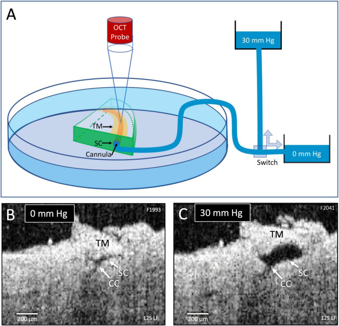
FIGURE 1.
Experimental setup and real-time pressure-dependent optical coherence tomography (OCT) images. (A) A tissue wedge in a Petri dish with the trabecular meshwork (TM) facing upward during high-resolution OCT imaging. A micromanipulator guides one end of a cannula to the wedge and holds it securely in Schlemm’s canal (SC). The opposite end of the cannula leads to a valve that switches between two reservoirs, one at 0 mm Hg and the other at 30 mm Hg. OCT imaging is initiated before switching reservoirs and continues until achieving a new steady-state tissue configuration. (B,C) OCT images at 0 and 30 mm Hg, respectively, from the distal SC location, are noted in Figure 2. Both SC and collector channels (CC) collapse at 0 mm Hg but markedly enlarge when pressure increases in SC.

