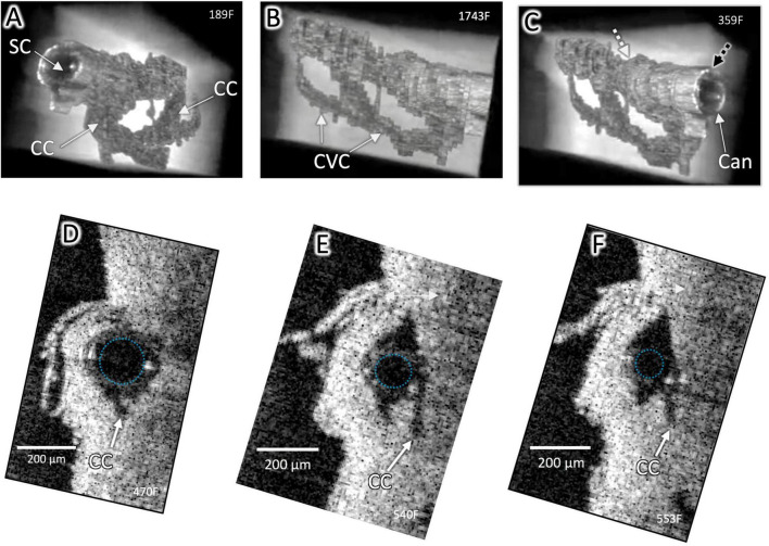
FIGURE 2.
Surface architecture and internal structure with a cannula in Schlemm’s canal. High-resolution OCT imaging permits delineation of the lumen boundaries of the outflow system after segmentation to eliminate surrounding structures in individual OCT B-scans and reconstruction with Amira 3D volumetric software. A cannula (Can) is visible in SC in (A–C), and the cannula’s internal diameter is outlined in (D–F). CC connect with an intrascleral circumferential-oriented vascular compartment (CVC) adjacent to and parallel with SC. Cannula location in (C) (Black dashed arrow) shown in cross-section in (D). Location of end of cannula denoted by the white arrow in (C) and cross-section in (F). The striped arrow distal to the cannula in (C) is the location of Figure 1 (B,C). The outside diameter (OD) of the cannula at (C) was 241 μm and at (D) was 178 μm, typical of stent sizes and here occludes the CC entrance. Distally, the smaller cannula diameter no longer occludes CC. Both CC and CVC are distended distal and proximal to the cannula end, demonstrating a pathway for fluid entry to the distal system from the cannula end (Supplementary Video 1).

