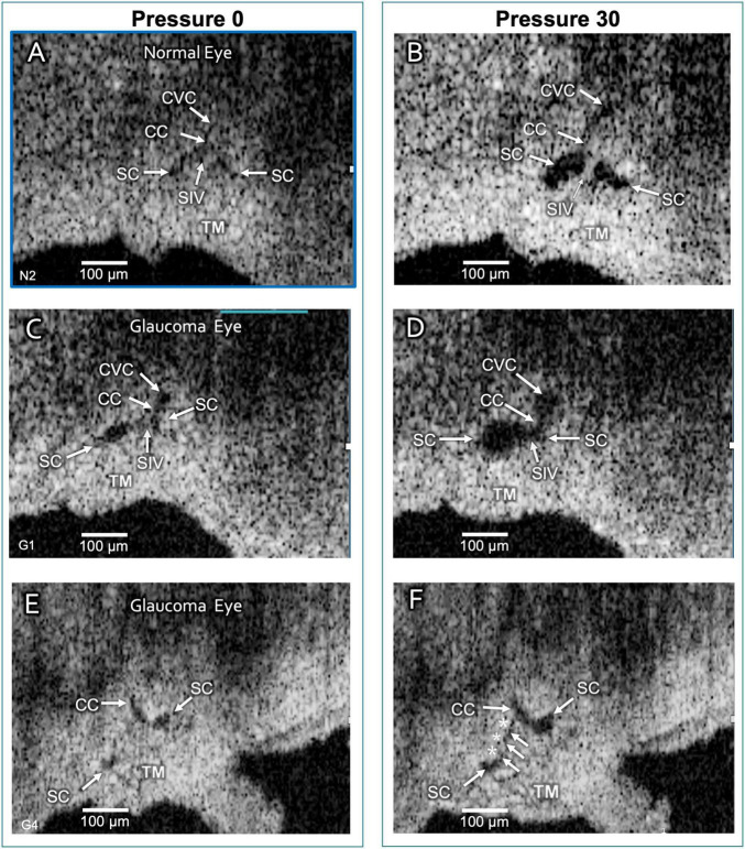
FIGURE 6.
Schlemm’s canal and collector channel configuration changes. The 2D high-resolution cross-section (i.e., B-scan) OCT images in (A,C,E) are at 0 mm Hg hydrostatic reservoir pressure, while images in (B,D,F) are at 30 mm Hg. Schlemm’s canal (SC) spans between the paired arrows. In (B,D), the lumen of SC, collector channel (CC) entrances, and the circumferentially-oriented vessel compartments (CVC) become markedly enlarged. Schlemm’s canal inlet valve-like structure (SIV) attachments between the trabecular meshwork (TM) and SC external wall elongation. (E,F) are from a glaucomatous eye. In (F), the TM remains in the CC entrance. TM motion is marked, but adhesions between the TM (white arrows) and SC external wall (asterisks) prevent SC from opening.

