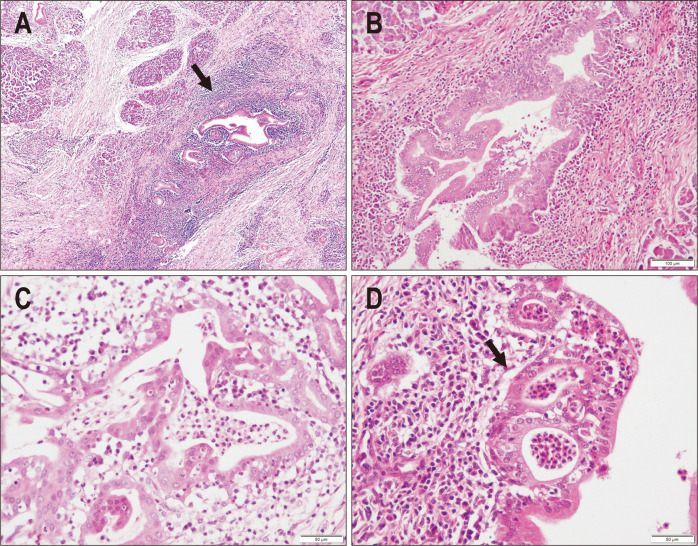
Fig. 1.
Microscopic features of type 2 autoimmune pancreatitis (AIP). (A) The pancreatic duct and lobules are both involved in the fibroinflammatory process, with the inflammatory infiltrate being more pronounced in the periductal area (arrow) (H&E, ×20). (B) The pancreatic duct is heavily infiltrated by inflammatory cells, including lymphocytes and plasma cells, and the architecture of the lining epithelium is irregular (H&E, ×40). (C) The pancreatic duct is severely damaged with many infiltrating neutrophils, which is consistent with granulocytic epithelial lesions (H&E, ×100). (D) Neutrophilic aggregates in the duct lumen resemble crypt abscesses in inflammatory bowel disease (H&E, ×100).

