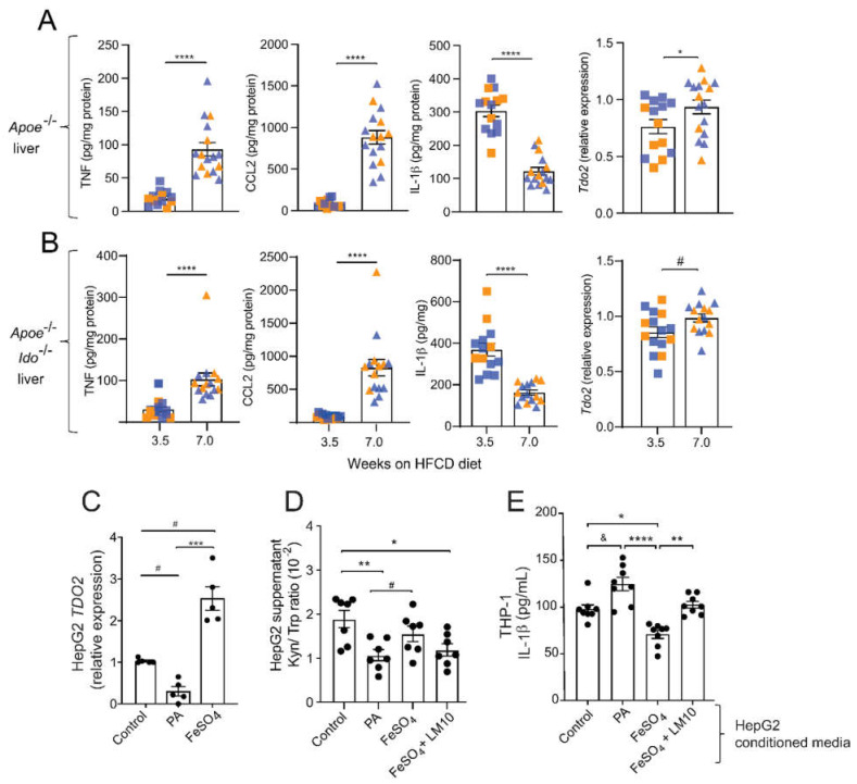
Figure 5.
Fatty acids and iron regulate TDO2-dependent Trp degradation and consequences for THP-1 macrophage activation. (A,B) TNF, CCL2, and IL-1β protein levels and relative Tdo2 mRNA expression in livers from Apoe−/− and Apoe−/−Ido1−/− mice fed a high-fat cholesterol diet (HFCD) for 3.5 and 7 weeks. (A,B) Pooled data from two independent experiments are shown; n = 14–15/group. © Relative expression of TDO2 mRNA in HepG2 cells treated with palmitic acid (PA, 500 μM) or iron (FeSO4, 100 μM) (n = 5). (D) Kyn/ Trp ratio in the supernatants of HepG2 cells treated with PA (500 μM), FeSO4, (100 μM), or FeSO4 + TDO2-inhibitor LM10 (0.62 μM) (n = 7). (E) IL-1β release from THP-1 macrophages pre-treated with conditioned media from HepG2 cells incubated with PA (500 μM), FeSO4 (100 μM), or FeSO4 + TDO2-inhibitor LM10 (0.62 μM); in addition, cells were stimulated with LPS (10 ng/mL, 4 h) and ATP (5 mM, 30 min) for activation of the inflammasome and IL-1β release (n = 8). & p < 0.06; # p < 0.08; * p < 0.05; ** p < 0.01; *** p < 0.001; **** p < 0.0001. (A) Differences were detected using the Mann–Whitney U test. (B,C) Differences were detected using a one-way ANOVA and Dunn’s post hoc test.

