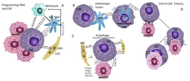
Figure 2.
Ecological behaviour of CSCs, autophagy and entosis. (A) Programming TAM and CAF. Activation of ERK/STAT3 signaling molecules triggers M2 polarization of monocytes in breast tumours [86]. The recruitment of monocytes from the blood is carried out due to the production of chemokines—CCL2, CCL18, and cytokines CSF1, VEGF, etc., by tumor cells. When polarizing monocytes in M2-TAM CSC, CSF1, HIF1a, TLR2, and other factors are used. [87]. CAF could promote monocyte recruitment toward cancer cells through CXCL12/CXCR4 pathway [88]. CAF produces MMPs that degrade the matrix and promote invasion of CSCs. CAFs produce IL17a which stimulates symmetric division of CSCs [89]. M2-TAMs produce IL4, 10, 13, TGFb, and PGE2, which act on CSCs and stimulate their symmetric division, increasing their population [90]. Thus, this type of relationship can be seen as a commensalism between TAM, CAF, and CSCs. The interaction between TAM and CSCs is described in detail in the overview [91]. (B) Heterotypic fusion. Heterotypic fusion cells expressed the tumour marker EpCam and macrophage markers CD163, CD68, CSFR1 and CD66b [92]. The hybrid cells display EMT with a significant downregulation of E-cad and upregulation of N-cad, Vim and Snail, as well as an obviously increased expression of MMP-2, MMP-9, uPA, and S100A4. The TCF/LEF transcription factor activity of the Wnt/β-catenin pathway and the expression of its downstream target genes including cyclin D1 and c-Myc increased in the hybrid cells [93]. This type of relationship can be viewed as a symbiosis with the acquisition of new properties that are not characteristic of individual populations. (C) Autophagy. With the induction of autophagy in CSCs, the expression of ATG5-12 LC3B, and BECLIN1, as well as the transcription factors FOXO3A, SOX2, NANOG, and STAT3, which can lead to the induction of the stem phenotype, increased resistance to chemotherapy, EMT, increased migration activity, and survival of dormant cells [94,95,96,97]. Under starvation and toxicity, CSCs can also induce autophagy of neighboring microenvironmental cells and non-CSCs, through exposure to ROS, IL6, and HIF1a. In this case, cells produce metabolites that feed CSCs, in particular lactate [94]. In this case, the behavior of CSCs can be seen as parasitism. (D) Cel-in-Cell. E-cadherin and P-cadherin are key components of the adherent junctions of cells in entosis process [98]. These molecules bind CSCs and non-CSCs scavenged. Increased RHO–RHO-associated coiled-coil-containing protein kinase (ROCK) and/or diaphanous-related formin 1 (DIA 1)–actomyosin show in losers cells [99]. CSCs express ezrin, actin, and caveolins, which mediate uptake [100]. Absorbed cells undergo apoptosis, autophagy, or are destroyed by lysosomal digestion, providing CSCs with nutrients [99]. In addition, entosis can lead to the formation of aneuploidy in CSCs [101], and this may be a rapid mechanism for the formation of new chemoresistant clones during treatment.

