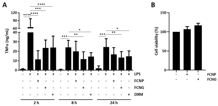
Figure 3.
Anti-inflammatory effect and toxicity of FCNP and FCNG in LPS-stimulated THP-1 macrophages (THP-1-MoM). (A) The evaluation of the inflammatory marker TNFα was performed by ELISA in the cell culture media of THP-1 MoM treated for 2 h, 8 h or 24 h with (11.7 ± 4.5) × 109 particles/mL of FCNP or FCNG, and then stimulated with LPS (100 ng/mL) for a further 24 h. Dexametasone (DXM) (2 μM) was used as a positive anti-inflammatory control and non-stimulated cells were used as controls for LPS stimulation. (B) Viability of THP-1-MoM exposed to (11.7 ± 4.5) × 109 particles/mL of FCNP or FCNG for 48 h. Data are representative of three independent experiments, and presented as mean ± SD. Two-way ANOVA and multiple comparisons were achieved with the Dunnett’s test and are presented relative to the LPS-stimulated cells. Statistical significance was defined as p ≤ 0.05 (*), p ≤ 0.01 (**), p ≤ 0.001 (***) and p ≤ 0.0001 (****).

