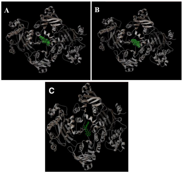
Figure 6.
A 3D representation of the binding of verapamil, curine, and guattegaumerine on the LmrA protein. The pictures presented in the figure are a graphical output of AutoDock 4.2. All of the representations of LmrA (ribbon in gray) are in the same orientation. (A) Verapamil on LmrA; (B) curine on LmrA; (C) guattegaumerine on LmrA. Verapamil and curine are represented in terms of their volume (green) and guattegaumerine is shown in a stick representation.

