Figure 7.
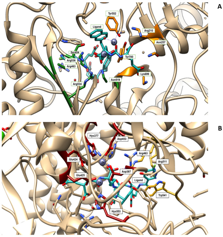
(A) Amino acids of the active site of the PSMA enzyme (GCPII) interacting with the ligand PSMA 1007 (PDB code 5O5T, in light-blue). S1 pocket (‘’non-pharmacophore pocket’’) with its arginine patch is indicated in green. It is specific for binding to the NAA (N-acetyl-aspartyl-) portion of NAAG (or the NAA-like- portion of PSMA-i) through polar or nonpolar interaction [112]. In orange is highlighted the S1′ pocket, which specifically binds C-terminal glutamate residue. S1 is a flexible funnel specific for negatively charged amino acids which improve the interaction between PSMA and their inhibitors; its flexibility enables the binding of a variety of groups, which are not essential to the determination of the affinity [113]. (B) The active site of the PSMA receptor in which amino acids deputy to stabilize zinc ions are colored in red, whereas the side chains of Arg463, Arg511 and Trp54, forming the “arene-binding site”, are colored in yellow. The latter define the entrance of GCPII [114]. The images are created using UCSF Chimera 1.14.

