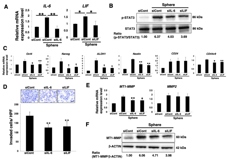
Figure 4.
Autocrine/paracrine IL-6 or LIF/gp130/STAT3 pathways involved in stemness and invasion of PDAC sphere cells. PANC-1 cells were cultured for 7 days in 2D (adherent) or 3D (sphere) conditions after siRNA transfection. The cells were harvested and then used in the following experiments. (A) Real-time qPCR analysis of IL-6 or LIF. Results are normalized to values obtained for adherent cells (value = 1). Results are presented as means ± SD from three independent experiments. (B) Western blot analysis of p-STAT3 and STAT3. Relative band intensity is presented. (C) Real-time qPCR analysis of stemness markers. Results are normalized to values obtained for adherent cells (value = 1). Results are presented as means ± SD from three independent experiments (* p < 0.05, ** p < 0.01 vs. siCont transfected sphere cells). (D) Matrigel invasion assays were performed in PANC-1 sphere cells. Representative results from measurements of 12 fields are presented (* p < 0.05, ** p < 0.01 vs. siCont transfected sphere cells). Scale bar: 100 μm. (E) Real-time qPCR analysis of MT1-MMP and MMP2. Results are normalized to values obtained for adherent cells (value = 1). Results are presented as means ± SD from three independent experiments (* p < 0.05, ** p < 0.01 vs. siCont transfected sphere cells). (F) Western blot analysis of MT1-MMP and β-ACTIN. Relative band intensity is presented.

