Figure 4.
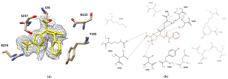
Active site of CTX-M-15 of the ampicillin-enzyme complex. (a) Electron density map of the CTX-M-15 active site bound to ampicillin in the yellow. 2Fo-Fc map (contoured at 1.0 sigma) is shown in dark gray. Residues around the binding pocket of the ampicillin -CTX-M-15 crystal structure are depicted as cream, red, blue sticks. The figure was generated with UCSF Chimera v1.13.1. (San Francisco, CA, USA). (b) 2D structure of ampicillin (orange) in the binding pocket of CTX-M-15. All residues have been shown, hydrogen bonds are drawn using dashed lines. The residues that interacted with ampicillin are shown in dark black, residues without interaction are depicted in gray. The figure was built by the MarvinSketch v.17.28.0. (Budapest, Hungary).

