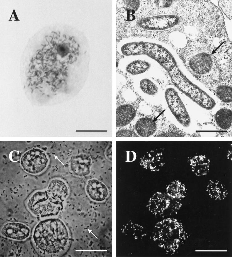
FIG. 2.
(A) Acanthamoeba trophozoites (UWC8 isolate) naturally infected with rod-shaped bacterial endosymbionts as seen with Hemacolor stain (bar, 7 μm). (B) Electron micrograph demonstrating intracellular bacterial symbionts (UWC8 isolate) and several mitochondria (arrows) in an Acanthamoeba trophozoite (bar, 1 μm). (C) Phase-contrast photomicrograph of fixed Acanthamoeba strain UWC8 trophozoites, with numerous E. coli food bacteria seen in the background (arrows) (bar, 15 μm). (D) Specific fluorescent in situ detection of the endosymbionts of Acanthamoeba strain UWC8 within the same field as seen in panel C; numerous rod-shaped intracellular bacteria are recognized by using probe AcRic1196 labelled with Cy3 (bar, 15 μm).

