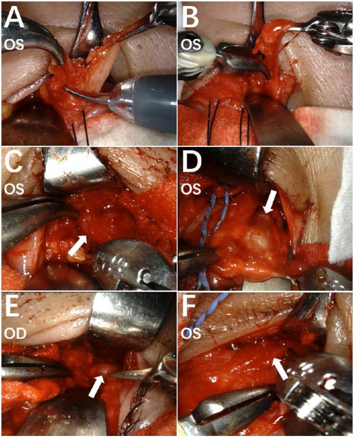
Figure 4.

The intraoperative view of the surgical field by the endoscope. Orbital fat was excised by monopolar curved scissor (A), grasped by a curved bipolar dissector, and separated by Black Diamond micro-forceps (B) to accomplish full removal. The extraocular muscles were clearly identified and protected during the operation, including inferior rectus muscle (C, white arrow), medial rectus muscle (D, white arrow), lateral rectus muscle (E, white arrow), and inferior oblique muscle (F, white arrow).
