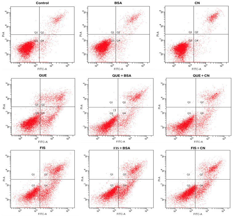
Figure 5.
Identification of the normal and apoptotic cells using the Annexin V/propidium iodide (PI) staining and flow cytometry. The cells were treated with 0.1% DMSO (control), quercetin (QUE), fisetin (FIS), bovine serum albumin (BSA), casein (CN), and the polyphenol–protein mixtures. The polyphenol dose was 80 μmol/L, while the protein dose was 1.0 g/L.

