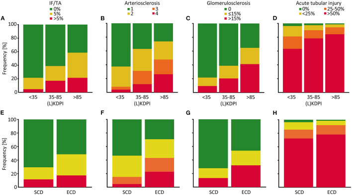
Figure 3.
Distribution of histological properties of post-reperfusion biopsies depending on the (L)KDPI of kidney transplantations after living and deceased donation. Percent stacked column chart of the amount of interstitial fibrosis and tubular atrophy (IF/TA), arteriosclerosis, glomerulosclerosis and acute tubular injury subdivided into (Living) Kidney Donor Profile Index [(L)KDPI] <35, 35–85, and >85% (A–D) or Standard Criteria Donor (SCD) and Expanded Criteria Donor (ECD) (E–H). Kidney graft tissue was taken 10 min after the onset of reperfusion by 18G core needle biopsy. Histological evaluation was performed by one renal pathologist blinded for clinical data. A semi-quantitative score according to the Banff Classification was used to assess arteriosclerosis. IF/TA, glomerulosclerosis, and acute tubular injury are shown as percentage of the entire area used for histological investigation.

