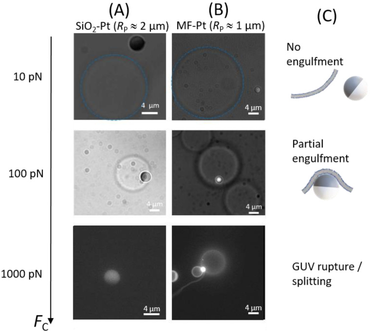
Figure 3.
Bright field and fluorescence microscopy images of: (A) SiO2–Pt Janus colloids (RP ≈ 2 µm) and (B) MF–Pt Janus colloids (RP ≈ 1 µm) interacting with POPC GUVs at given centrifugal forces FC. Images are taken from below. Dashed blue circles are guides to identify the vesicles. (C) Sketches of a Janus colloid non-interacting with a membrane at FC ≈ 10 pN, and a Janus colloid partially engulfed by a GUV membrane at FC ≈ 100 pN. For FC ≈ 1000 pN, the number and the size of GUVs dramatically decreased due to GUV rupture and splitting.

