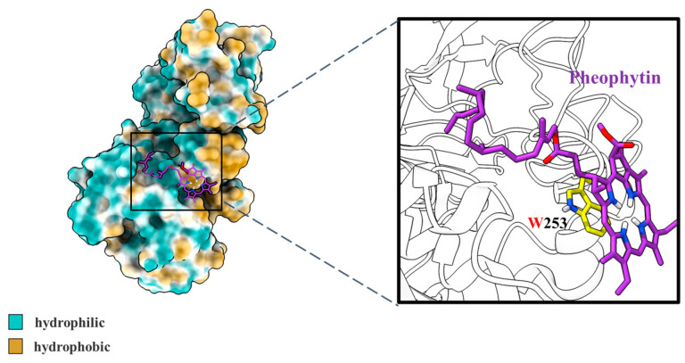
Figure 7.
Conformational analysis of molecular docking results for pheophytin and pancreatic lipase. The structure of pheophytin was drawn as sticks, while the structure of lipase was drawn as the surface. The hydrophilic and hydrophobic groups of pancreatic lipase were painted cyan and yellow, respectively. The solid frame area presented a close-up view of the binding site for pancreatic lipase and pheophytin.

