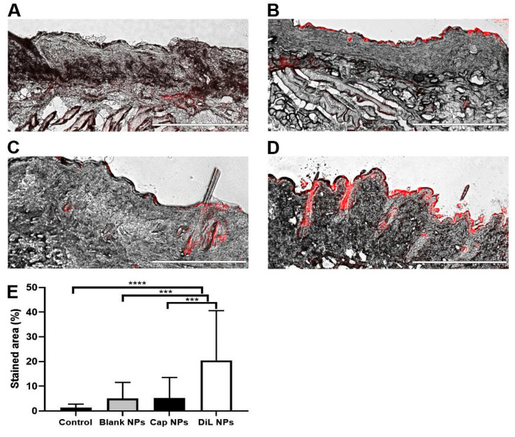
Figure 2.
Intracutaneous penetration of NPs topically applied to the skin in the mouse. Exemplary microscope images (Scale bar is 400 µm) of (A) a potential autofluorescence (red area) of untreated skin from mouse cheek, (B) after treatment with blank NPs, (C) after treatment with capsaicin NPs, (D) treated with red dye (red area) containing DiL NPs. (E) Mean values of red color measured in cheek tissue from 18 samples per group. *** p < 0.001, **** p < 0.0001, mean ± SD, one-way ANOVA with Bonferroni correction.

