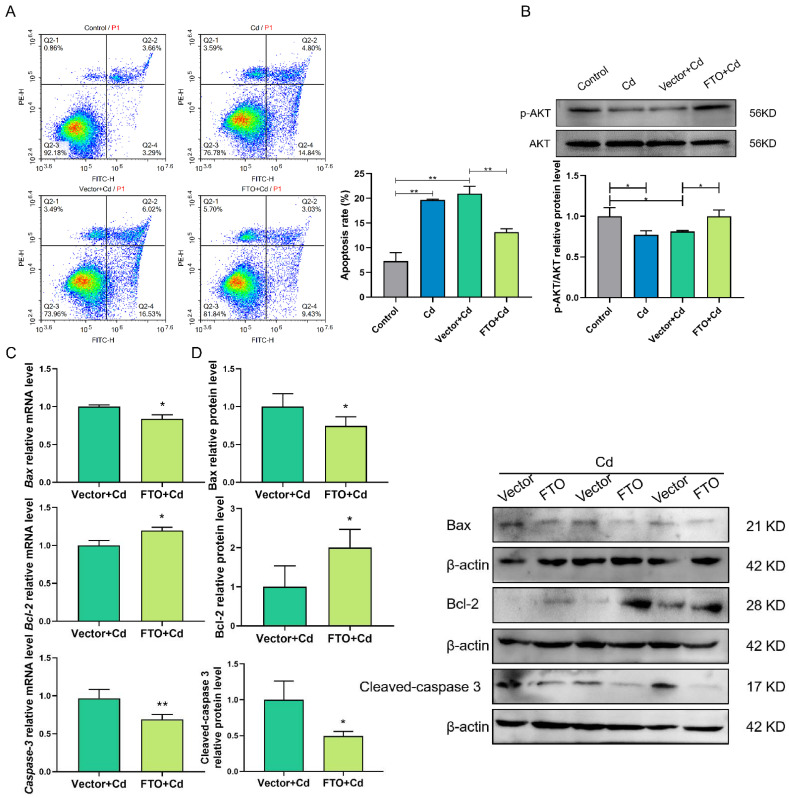
Figure 6.
FTO overexpression suppressed Cd-induced apoptosis, perhaps by activating the AKT pathway in granulosa cells. (A) Flow cytometry analysis results detected apoptosis rate in the control, Cd, Vector + Cd, and FTO + Cd groups. (B) The p-AKT protein levels were analyzed by Western blotting after Cd and FTO treatments. (C,D) RT-qPCR and Western blotting results indicated the mRNA and protein levels of Bax, Bcl-2, and caspase-3 after FTO overexpression. * p < 0.05; ** p < 0.01.

