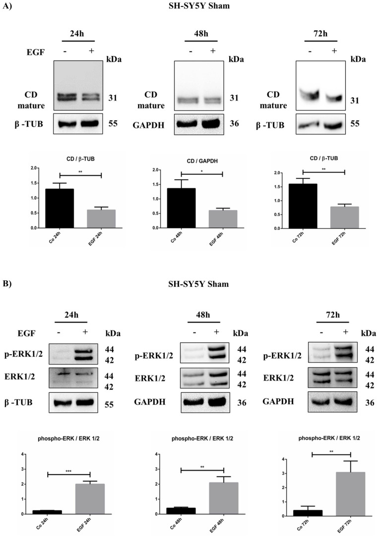
Figure 7.
EGF downregulates intracellular cathepsin D in SH-SY5Y Sham. SH-SY5Y cells were treated for 24, 48, and 72 h. (A) Time course of cathepsin D expression analyzed through western blotting. (B) Western blot of phospho-ERK 1/2 and total protein in Sham cell homogenates at different time points (24, 48, and 72 h) in the presence/absence of EGF. The filters were probed with β-tubulin and GAPDH as loading control. Significance was considered as follows: *** p < 0.001; ** p < 0.01; * p < 0.05.

