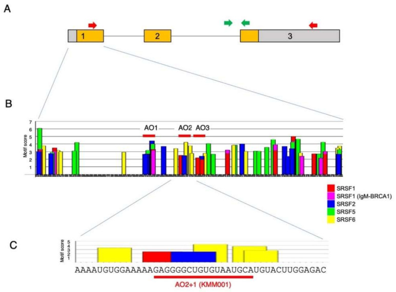
Figure 1.
Structure of MSTN pre-mRNA and exonic splicing enhancer sequences in exon 1. (A) Structure of MSTN pre-mRNA. The structure of MSTN pre-mRNA is schematically described. The MSTN gene comprises three exons and two introns. Exon 1 is 506 nt, and the ATG start codon is present at nucleotide 134. Boxes and lines indicate exons and introns, respectively. The number in the box indicates exon number. The colored area within the box indicates the myostatin-coding sequence. For detection of mature mRNA, a fragment extending from exon 1 to 3 (2328 bp) was PCR amplified using primers on the respective exons (red arrow). To determine the expression of MSTN pre-mRNA, a fragment extending from intron 2 to exon 3 (365 bp) was PCR amplified using primers for the respective regions (green arrow). (B) Exonic splicing enhancer sequences in exon 1 of the MSTN gene. The nucleotide sequence of exon 1 of the MSTN gene was analyzed for the presence of splicing enhancer sequences with ESEfinder3.0. A graph of MSTN exon 1 produced by ESEfinder3.0 is shown. Only the high-score values (above the selected threshold) are mapped on the output graph. For the color-coded bars, the height of the bar represents the motif score, the width of the bar indicates the length of the motif, and the color of the bar indicates the SR protein (SFRF1, SFRF1 (IgM-BRCA1), SFRF2, SFRF5, and SFRF6 are indicated by red, purple, blue, green, and yellow bars, respectively). Three ASOs (AO1, AO2, and AO3) were synthesized in the first step of screening (red bar). (C) Enlarged graph of the areas around sequences complementary to AO2+1 (KMM001). A part of the output graph (B) is shown. The bars and their coloring are identical to those in (B).

