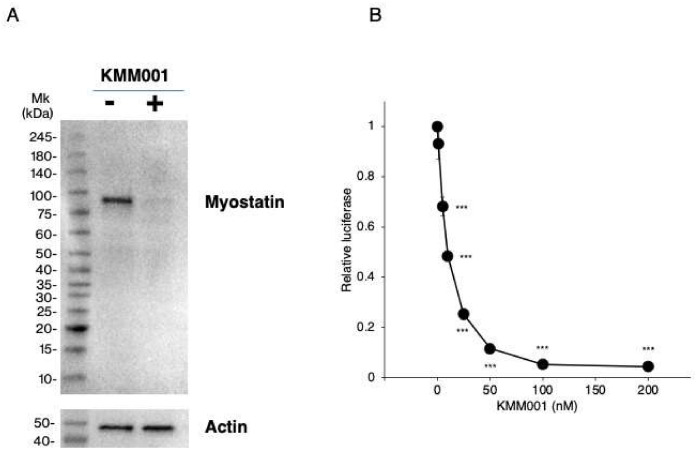
Figure 7.
Inhibition of myostatin protein expression and myostatin signaling by KMM001. (A). Myostatin protein levels in CRL−2061 cells transfected with AO2+1 were assayed by Western blot analysis using an antibody against the N-terminal domain of human myostatin. SDS–PAGE was carried out under nonreducing conditions. Immunoblotting results are shown. One clear band was identified in untreated CRL−2061 cells (left lane). In contrast, in KMM001-treated cells, a band corresponding to myostatin was weakly visualized (right lane). Mk refers to the size marker. (B). Myostatin signaling was assayed in CRL−2061 cells using the SMAD-dependent luciferase reporter gene. The reporter gene was transfected into cells together with a galactosidase plasmid as a control. The luciferase activity was normalized to the galactosidase activity, and the ratio of luciferase activity to galactosidase activity was calculated. The relative luciferase activity was set to 1 in the nontreated cells. The relative luciferase activity decreased dose dependently from 0 to 100 nM KMM001 and reached a plateau. *** = p < 0.001 vs. 0 nM KMM001.

