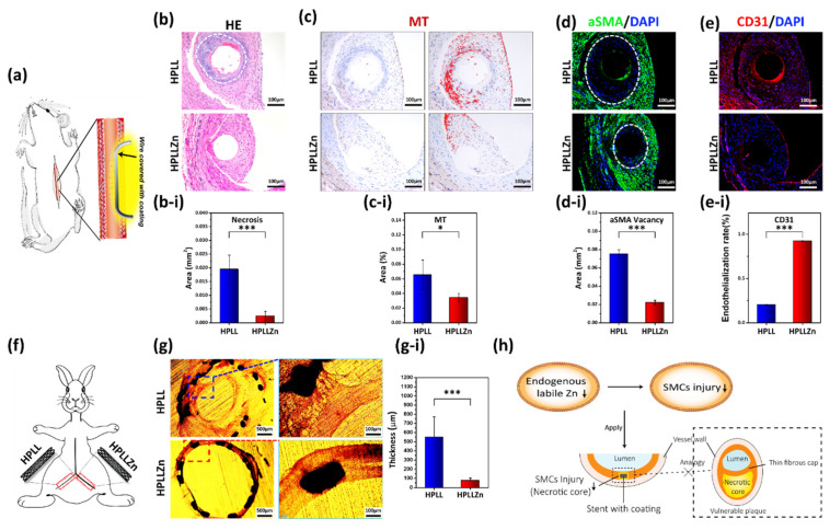
Figure 5.
(a) Schematics of stainless-steel wires with different coatings implanted into Sprague Dawley rats’ abdominal aorta. (b) Hematoxylin and eosin staining and (b-i) quantification of the necrotic area surrounding the different coatings (the white dotted line shows the necrotic area). (c) Immunohistochemical of metallothionein protein expression and (c-i) semiquantitative analysis of the area (red marks regions with high MT expression). (d) The expression edge of α-SMA (green) and (d-i) quantification of the area without αSMA in the tissue close to the coating after a 1-month implantation (the white dotted line shows the edge closest to the coating). (e) Immunofluorescence staining of CD31 and (e-i) the endothelialization integrity. (f) Schematics of vascular stents with different coatings implanted in New Zealand white rabbits’ iliac artery. (g) Histomorphometric analysis of stents after a 1-month implantation and (g-i) quantification of the mean neointimal thickness. (h) The potential mechanism of this rule applies to the cardiovascular stent. (HE, hematoxylin and eosin stain; MT, metallothionein; α-SMA, α-smooth muscle actin; CD31, cluster of differentiation 31; DAPI, 2-(4-Amidinophenyl)-6-indolecarbamidine dihydrochloride; data were analyzed using 1-way ANOVA, * p < 0.05, *** p < 0.001; ns, not significant).

