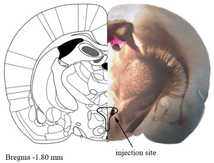
Figure 1.
A representative photograph of the microinjection site in the paraventricular nucleus of hypothalamus (PVN) coupled with the matching slide from the Paxinos and Watson rat brain atlas [24]. One mm thick brain slice with the injection site shown by Evans blue dye. The drawn outline represents the confines of PVN.

