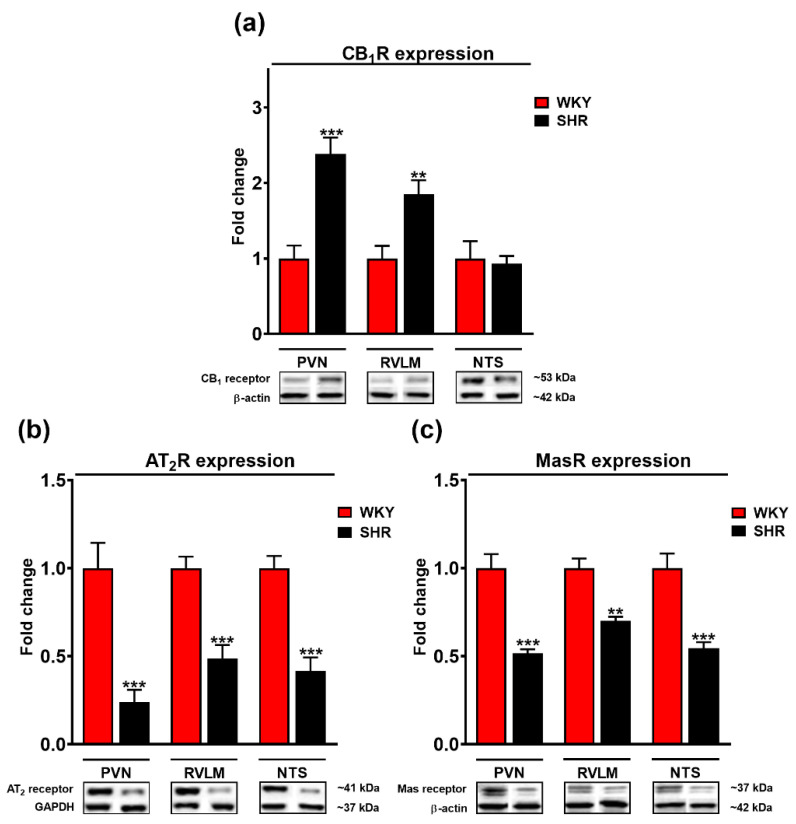
Figure 7.
Fold change of cannabinoid CB1 (a), AT2 (b) and Mas (c) receptors and representative Western blots of the paraventricular nucleus of hypothalamus (PVN), rostral ventrolateral medulla (RVLM) and nucleus tractus solitarii (NTS) in Wistar Kyoto (WKY) and spontaneously hypertensive rats (SHR). β-actin or glyceraldehyde-3-phosphate dehydrogenase (GAPDH) served as loading control. Results presented as mean ± SEM (n = 6). ** p < 0.01, *** p < 0.001 compared to WKY. Uncropped Western blot images can be seen in Supplementary Materials, file Figure S1.

